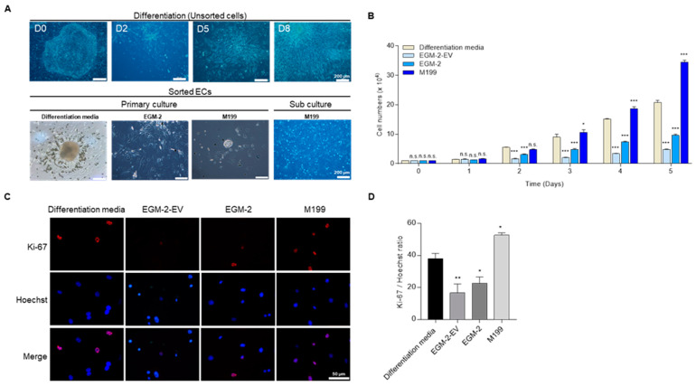
Figure 2.
The proliferation of sorted ECs. (A) Morphologies of pEpiSCs and differentiated cells in differentiation media on the days 0, 2, 5, 8, respectively. Sorted ECs were cultured in differentiation media, EGM-2, or M199 culture system on 0.2% gelatin for the primary culture. Primary sorted ECs cultured in differentiation media were transferred to M199 culture system for sub-culture. (B) Proliferation rates of 1.0 × 10⁴ of sorted ECs were evaluated in four culture conditions. Values presented as mean SEM. * p < 0.05, ** p < 0.01, *** p < 0.001 vs. differentiation media, n.s.: not significant. (C) Immunofluorescence of Ki-67 in differentiation media, EBM-2-EV, EGM-2 and M199. Red: staining of Ki-67, blue: staining of Hoechst. Scale bar = 50 μm (D) Quantification of Ki-67 positive cells in culture of sorted ECs in differentiation media, EBM-2-EV, EGM-2 and M199. Values presented as mean SEM. * p < 0.05, ** p < 0.01 vs. differentiation media, n.s.: not significant.

