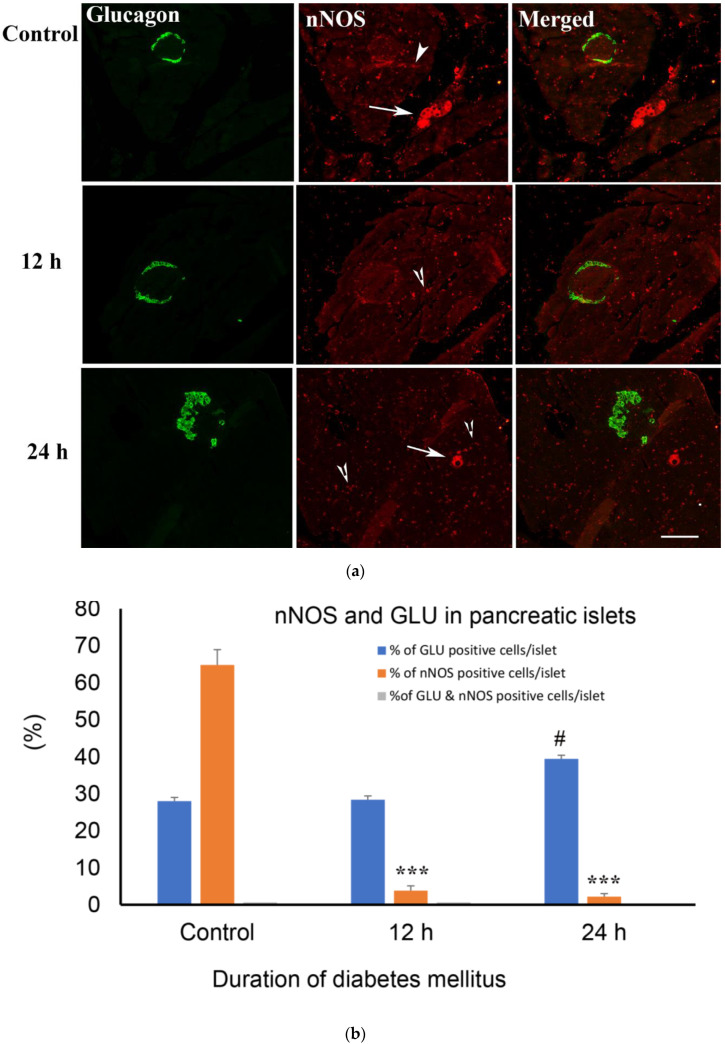
Figure 6.
(a) shows glucagon (GLU)-(green) and nNOS-(red)-immuno-positive cells in the islet of Langerhans of rats of control and those with 12 h and 24 h of diabetes. There was no co-localization between glucagon and nNOS. nNOS-immunoreactive ganglion cell (arrow) and varicose nerve fibres (arrow head) were observed in the exocrine pancreas. Note the significant increase in the number of nNOS-containing nerve profiles after 12 and 24 h of diabetes. Scale bar = 10 μm. (b) shows morphometric analysis of glucagon-positive cells in the islet cells of the endocrine pancreas. There was a significant (*** p< 0.001) reduction in the number of nNOS-immunoreative cells in the islet of Langerhans compared to control. The number of glucagon-positive cells in the islets increased markedly (# p < 0.01) 24 h after the induction of diabetes, compared to the control (n = 6).

