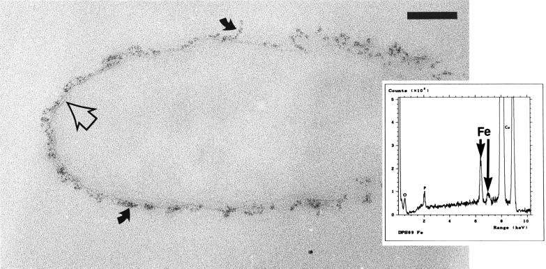
FIG. 5.
Thin section of an iron-treated strain dps89 cell. The iron bound to such an extent that membranes were sometimes visible (open arrow), and amorphous precipitates (solid arrows) formed on the cell surface. Bar = 100 nm. (Inset) EDS spectra of the precipitates confirmed that they were iron rich. The lack of other high-atomic-number elements suggested that the precipitate might be an iron hydroxide. These results were obtained only for strain dps89.

