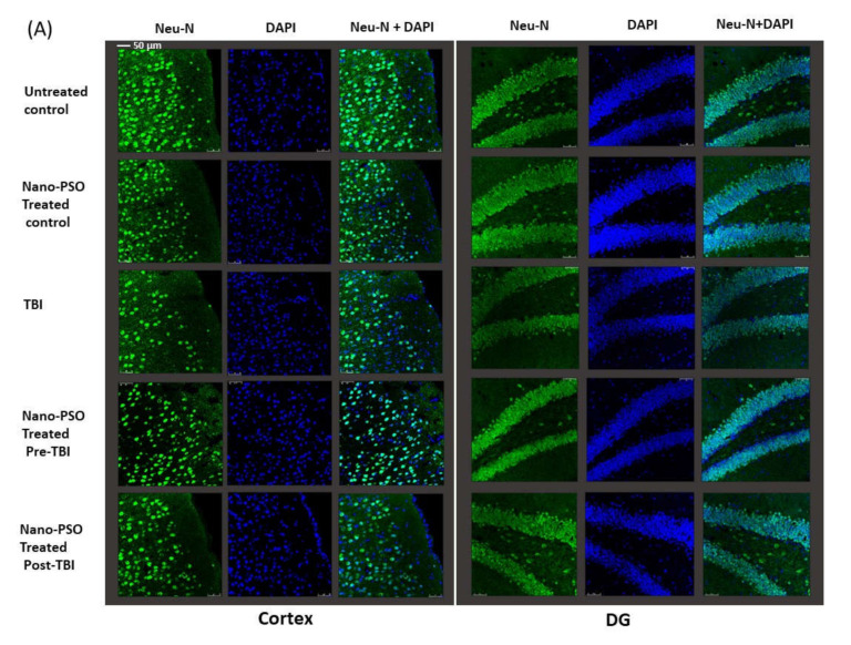
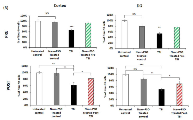
Figure 4.
Nano-PSO treatment mitigates neuronal loss in the brain of TBI mice: (A) coronal sections through the temporal cortex and dentate gyrus of brains from treated and untreated controls, as well as from untreated TBI, treated with Nano-PSO before TBI and treated with Nano-PSO post-TBI were immunostained for NeuN (green) and DAPI (blue) [scale bar 50 µm]; (B) quantitative assessment of NeuN immunostaining for all sections (bregma temporal cortex −1.06 nm and dentate gyrus −1.34 nm) at the end point of each experiment was quantified using One-way ANOVA (Cortex: PRE (F (3, 16) = 15.469, p = 0.000 Fisher’s LSD post hoc, *** p < 0.001, N = 5); PSOT (F (3, 12) = 8.607, p = 0.000 Fisher’s LSD post hoc, * p < 0.05 ** p < 0.01, n = 3–5)), (DG: PRE (F (3, 14) = 6.804, p = 0.000 Fisher’s LSD post hoc, ** p < 0.01, n = 4–5); PSOT (F (3, 13) = 12.580, p = 0.000 Fisher’s LSD post hoc, * p < 0.05, ** p < 0.01, *** p < 0.001, n = 3–4)). NS = Not significant. Values are presented as mean ± SEM.


