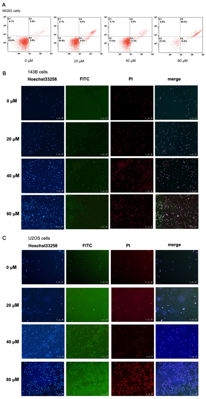
Figure 3.
Effect of α-linolenic acid on the apoptosis of MG63 osteosarcoma cells, and 143B and U2OS cells. (A) MG63 cell apoptosis was analyzed by flow cytometry with Annexin V and PI double staining. The four regions represent the state of cells: Alive, necrosis, early apoptosis, and late apoptosis. (B,C) Hoechst 33258 staining and Annexin V/PI double staining were performed in 143B and U2OS cells treated with α-linolenic acid, respectively. The concentrations of α-linolenic acid were 0, 20, 40, and 80 μM.

