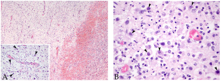
Figure 1.
Brain cerebral cortex sample of case 4, a male striped dolphin calf presenting 7/7 of the evaluated brain lesions. (A) Suffusive haemorrhages and malacia are present. Original magnification ×10; haematoxylin and eosin staining. Inset: numerous basophilic intranuclear inclusion bodies were observed within the neurons and glial cells (arrowheads). Original magnification ×40; haematoxylin and eosin staining. (B) Higher magnification of the basophilic intranuclear inclusion bodies (arrowheads). Original magnification ×60; haematoxylin and eosin staining.

