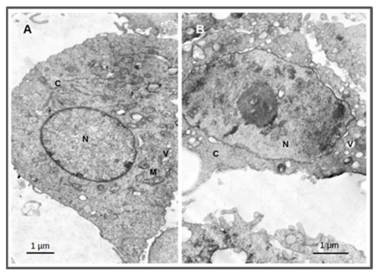
Figure 10.
Thin-section transmission electron microscopy images of PC3 cells treated for 4 h with AuNP-BBN at a concentration of 36 µg Au/mL followed by irradiation with 2 Gy (1 Gy/min). Samples were analyzed and photographed in a JEOL 1200-EX electron microscope. Irradiated PC3 cells (A); irradiated PC3 cells after incubation with AuNP-BBN. (B); N = nucleus; C = cytoplasm; M = mitochondria; V = vacuoles. Bars = 1 micrometer.

