Figure 5.
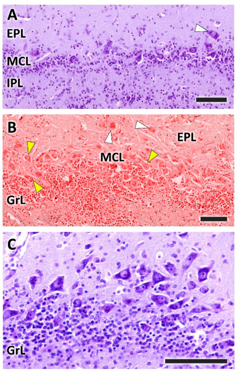
Histological study of the mitral cell layer of the olfactory bulb of the dog. (A,B) Nissl and Tolivia staining, respectively, of the mitral cell layer (MCL) showing typical mitral cells (yellow arrowheads) and deep tufted cells (white arrowheads) scattered along the boundaries between the external (EPL) and internal plexiform layers (IPL). (B,C) Sagittal section of the OB reveals a higher density of mitral cells, which are mitered or triangular in shape. Tolivia and Nissl staining, respectively. Scale bars: (A,B): 250 µm; (C): 100 µm.

