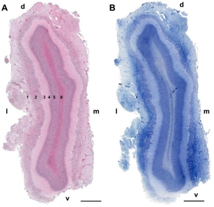
Figure 10.
Transverse section of the olfactory bulb of the wolf stained by hematoxylin–eosin (A) and Nissl staining (B). From superficial to deep, the following layers are identified: (1) Olfactory nerve layer (NL); (2) Glomerular layer (GlL); (3) External plexiform layer (EPL); (4) Mitral cell layer (ML); (5) Granular layer (GrL); (6) White matter: d, dorsal; l, lateral; m, medial; v, ventral. Scale bars: 2 mm.

