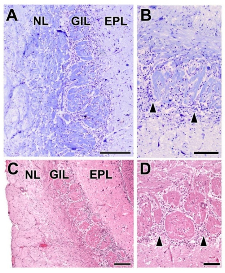
Figure 12.
Histological study of the superficial layers of the olfactory bulb of the wolf. (A,B) Nissl staining of the nerve (NL), glomerular (GlL) and external plexiform (EPL) layers. The arrowheads indicate the presence of periglomerular cells. (C,D) Hematoxylin–eosin staining. The periglomerular cells are mainly located in the border between the glomeruli and the EPL (arrowheads). Scale bars: (A): 250 µm; (B): 125 µm; (C,D): 100 µm.

