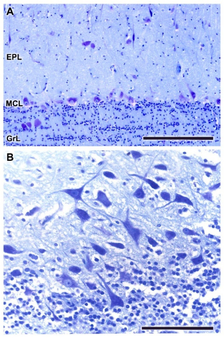
Figure 20.
Histological study of the mitral cell layer of the olfactory bulb of the fox. (A) Nissl staining of a transverse section of the olfactory bulb showing mitral cell somas aligned along the MCL. (B) A sagittal section of the mitral cell layer (MCL) stained with Nissl staining shows the typical mitral cells characterized by their big soma and well-defined dendritic projections. Scale bars: (A): 250 µm. (B): 100 µm.

