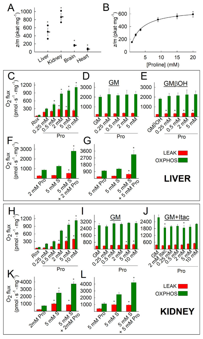
Figure 1.
Kinetic characterization of ProDH activity in isolated mitochondria (A,B), and the effect of proline concentration on oxygen consumption rates (LEAK: red bars; OXPHOS: green bars) of isolated mouse liver (C–G) and kidney (H–L) mitochondria using various substrate combinations and concentrations. (A) ProDH catalytic activity content (expressed in pkat/mg) of mitochondria isolated from mouse liver, kidney, brain, and heart using saturating concentrations of proline (100 mM) * p < 0.05 (comparing all groups with each other; ANOVA on Ranks). Data are SEM averaged from three independent experiments. (B) Determination of apparent Km of mouse liver ProDH for proline. Data points are SEM averaged from three independent experiments. (C,H) Rox: residual oxygen consumption (no external substrate added; increased OXPHOS indicates the effect of ADP stimulating respiration on internal substrates), followed by proline titrations. (D,I) GM: glutamate and malate, followed by proline titrations. (E) GM and 2 mM βOH, followed by proline titration. (J) GM: glutamate and malate, 2 mM Itac: GM+2 mM itaconate, followed by proline titration. (F,K,G,L) Proline (Pro) and/or succinate (S). Data are SEM averaged from at least three independent experiments. For C–L, * p < 0.05 (ANOVA Bonferroni [comparisons are to controls, i.e., no proline] or on ranks, if normality failed).

