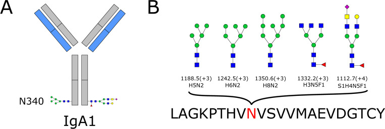
Fig 1.
A) IgA1 with N-glycosylation site 340 shown. B) The five different glycan compositions identified at site 340 of IgA1. Five different N-glycopeptides were identified that belonged to IgA1 and contained glycans attached at site 340. Fig 1A shows IgA1 with two of these glycans as an example (it is not possible to determine if these two glycans were both detected on the same IgA1 antibody). The heavy chains are shown in gray and the light chains in blue. The three high-mannose N-glycans and two complex-type N-glycans identified at site 340 are shown in Fig 1B. Monosaccharide symbols follow the SNFG (Symbol Nomenclature for Glycans) system [18]. The m/z value and charge are given below the glycan structures. For the glycan structures, the following abbreviations were used: H = hexose, N = N-acetylhexosamine, F = fucose, S = sialic acid. The peptide sequence is given at the bottom of the figure, with asparagine (N) being the amino acid to which the glycan is linked. L = leucine, A = alanine, G = glycine, K = lysine, P = proline, T = threonine, H = histidine, V = valine, S = serine, M = methionine, E = glutamic acid, D = aspartic acid, C = cysteine, Y = tyrosine.

