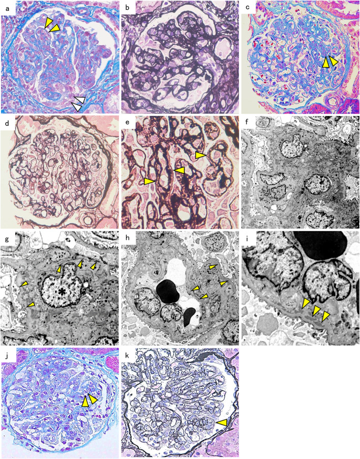
Fig. 1.
Kidney biopsy findings. a Light microscopy findings in case 1 show that the glomeruli have diffuse mesangial proliferation with endocapillary hypercellularity (yellow arrowheads) and cellular crescent (white arrowheads). Masson’s trichrome stain (× 400 original magnification). b Some glomeruli in case 2 show lobular patterns with the capillary wall thickening. Periodic acid methenamine silver (PAM) stain (× 400 original magnification). c Light microscopy findings in case 2 show glomeruli with endocapillary hypercellularity (yellow arrowheads). Elastica––Masson stain (× 400 original magnification). d, e The glomerular capillary walls in case 2 show duplication (yellow arrowheads). PAM stain (× 400, × 600 original magnification). f Electron microscopy findings in case 2 showing mesangial and paramesangial deposits (× 1200 original magnification) and g a double contour of the glomerular basement membrane (yellow arrowheads) with mesangial interposition (asterisk) (× 3000 original magnification) or h without mesangial interposition (× 1200 original magnification). i Subendothelial deposits (yellow arrowheads) (× 3000 original magnification) in case 2. j Light microscopy findings in case 3 show glomeruli with endocapillary hypercellularity (yellow arrowheads). Masson’s trichrome stain (× 400 original magnification). k The glomerular capillary walls in case 3 show duplication (yellow arrowhead). PAM stain (× 400 original magnification)

