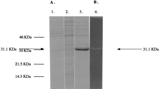
FIG. 4.
Expression of lyt51 in E. coli. Lane 1, SDS-PAGE of an extract (2.68 μg of protein) of E. coli cells containing plasmid pQE60 which were grown in the presence of an inducer (control); lane 2, SDS-PAGE of an extract (3 μg of protein) of cells harboring pMS51 which were grown in the absence of an inducer (lyt51 was not expressed); lane 3, SDS-PAGE of an extract (3.2 μg of protein) of pMS51-containing E. coli cells that were grown in the presence of inducer (lyt51 was expressed); lane 4, renaturing SDS-PAGE (with copolymerized crude cell walls of S. thermophilus CNRZ1205 as a substrate [see Materials and Methods]) of an E. coli cell extract (1.5 μg of protein) containing pMS51; the cleared area represents the position where the incorporated cell walls have been hydrolyzed through the action of a lytic activity. The sizes of the molecular mass markers are indicated on the left; the position of the lytic activity/Lyt51 protein is indicated by arrows.

