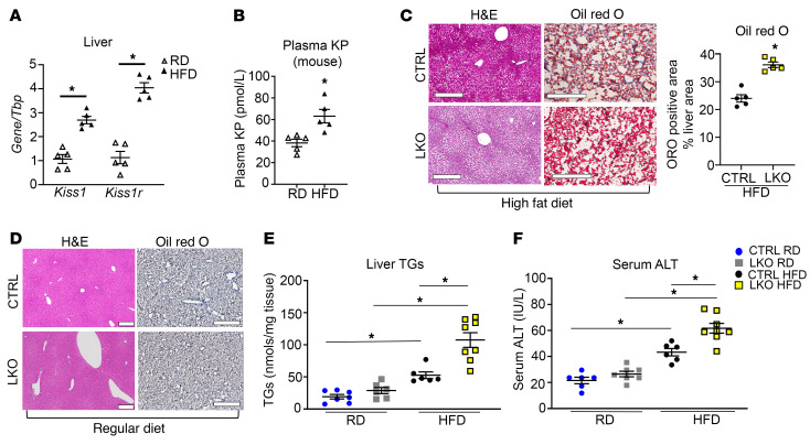
Figure 1. Hepatic Kiss1r-knockout mice exhibit increased hepatic steatosis in a diet-induced mouse model of NAFLD.
(A) Expression of Kiss1 and Kiss1r by RT-qPCR and (B) plasma KP levels in C57BL/6J male mice on regular diet (RD) or high-fat diet (HFD) for 12 weeks. (C and D) Representative histology of H&E- (showing steatosis) or Oil Red O–stained (showing lipids, red) liver sections. Quantification of staining is shown. Scale bars: 500 μm. No Oil Red O staining was observed in D. (E) Liver triglycerides (TGs) and (F) serum alanine aminotransferase (ALT) levels in control (CTRL) and hepatic Kiss1r-knockout (LKO) mice after 20 weeks on RD or HFD diet. Student’s unpaired t test or 1-way ANOVA followed by Dunnett’s post hoc test; *P < 0.05 versus respective controls.

