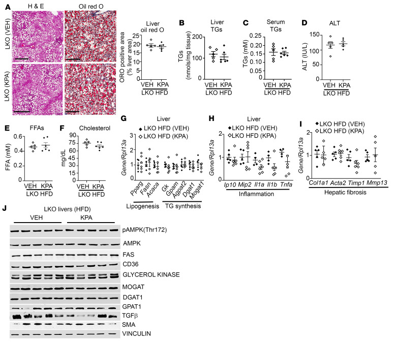
Figure 6. Lack of effect of kisspeptin analog treatment in hepatic Kiss1r-knockout mice fed HFD.
(A) Representative histology of H&E-stained liver sections showing steatosis (left) and Oil Red O–stained (right) liver sections showing lipid accumulation. Quantification of staining is shown. Scale bars: 200 μm. KPA, kisspeptin agonist. (B and C) Levels of (B) liver and (C) serum triglycerides (TGs). (D–F) Serum (D) ALT, (E) free fatty acids (FFA), and (F) cholesterol levels in hepatic Kiss1r-knockout (LKO) mice fed HFD for 11 weeks. (G–I) Expression of indicated genes by RT-qPCR. (J) Representative Western blots showing expression of indicated proteins. Densitometric analyses of blots is shown in Supplemental Figure 8, F–N. Data are shown as the mean ± SEM. Student’s unpaired t test; *P < 0.05 versus respective controls.

