Figure 4.
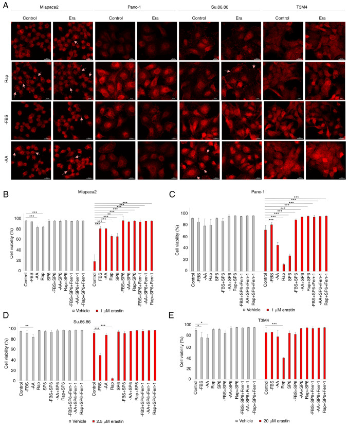
Starvation regulates ferroptosis via JNK. (A) Confocal microscopy images of phospho-JNK staining in (pseudo)starved Miapaca2, Panc-1, Su.86.86 and T3M4 cells, with or without erastin treatment. Arrowheads indicate phospho-JNK accumulation in the cell membrane. Scale bar, 20 μm. (B-E) Cell viability assessment of (B) Miapaca2, (C) Panc-1, (D) Su.86.86 and (E) T3M4 cells, cultured without FBS, without amino acids L-glutamine, L-lysine and L-arginine, pseudo-starved by treating with rapamycin and treated with the JNK inhibitor, SP6 (5 µM). Ferroptosis was induced by erastin and inhibited by Ferr-1. The data are presented as the mean ± SD; n=3. Cells cultured under standard conditions were used as the control. *P<0.05, **P<0.01 and ***P<0.001, vs. control. -AA, cells, cultured without L-glutamine, L-lysine and L-arginine; Rap, rapamycin; SP6, SP600125; Ferr-1, ferrostatin-1.

