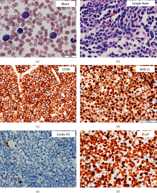
Figure 1.

(a) Wright–Giemsa stain of peripheral blood smear shows circulating large atypical lymphoid cells, highlighted with arrows (1000×). (b) The hematoxylin and eosin section shows diffuse proliferation of large pleomorphic lymphoid cells in the cervical lymph node (600×). (c–f) Immunohistochemical stains (all 600×) demonstrate that the cervical lymph node neoplastic cells are positive for CD20 and SOX-11, while negative for cyclin-D1. Ki-67 highlights a very high proliferative index of ∼90%.
