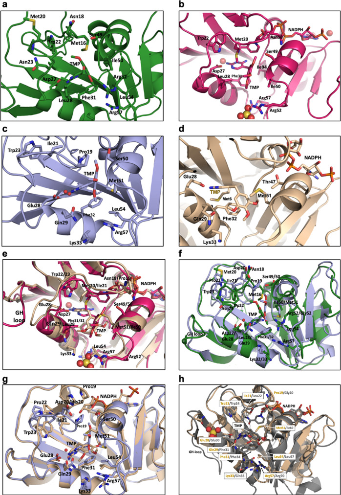
Fig. 3. Crystal structures of DHFR with trimethoprim (TMP).
a EcDHFR:TMP (green), b EcDHFR:TMP:NADPH (magenta), c DfrA1:TMP (light blue), and d DfrA5:TMP:NADPH (beige). e Overlays of the crystal structures of DHFR with TMP ternary complexes; EcDHFR:TMP:NADPH (magenta) superimposed onto DfrA5:TMP:NADPH (beige), f Binary complexes; EcDHFR:TMP (green) superimposed onto DfrA1:TMP (light blue), g DfrA1:TMP (light blue) and DfrA5:TMP:NADPH (beige), and h DfrA5:TMP:NADPH (beige) superimposed onto Human DHFR:TMP:NADPH (PDB id code 2W3A). Residues important to ligand binding are shown in stick mode and colored by atom.

