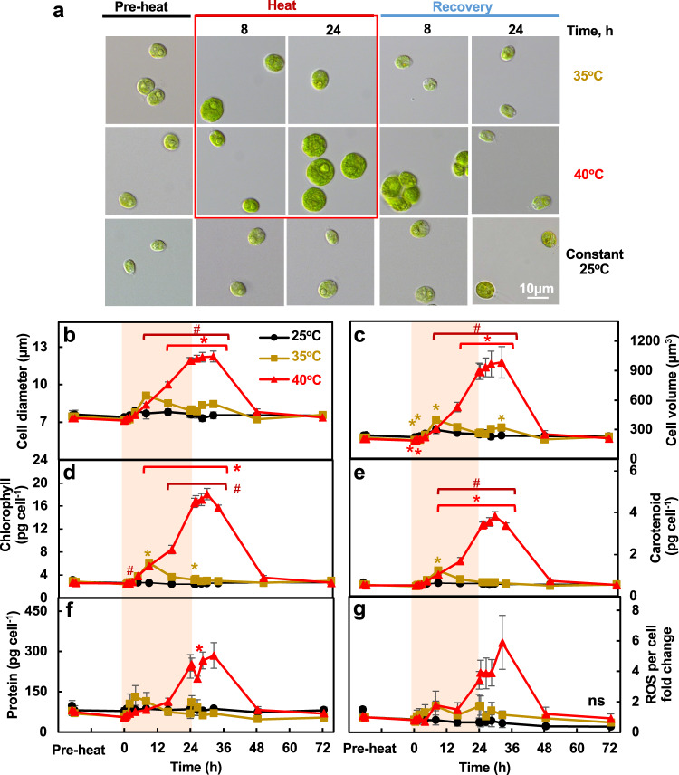
Fig. 5. Heat of 40°C persistently increased cell size, cellular levels of pigments, and proteins while these effects were transient with 35°C heat.
a Light microscopic images of Chlamydomonas cells. b, c Cell diameters and volume determined using a Coulter Counter. d–f Total chlorophyll, carotenoid, protein content per cell. g Fold-change of reactive oxygen species (ROS) levels per cell quantified using CM-H2DCFDA ROS indicator. Mean ± SE, n = 3 biological replicates. Black, brown and red curves represent experiments with constant 25°C, treatments of 35°C or 40°C respectively. Red shaded areas depict the duration of high temperature. Statistical analyses were performed using two-tailed t-test assuming unequal variance by comparing treated samples with 25°C at the same time point (*, p < 0.05) or between 35°C and 40°C at the same time point (#, p < 0.05). b–g P values were corrected by FDR. The colors and positions of asterisks (*) match the treatment conditions and time points, respectively. The positions of pound signs (#) match the time points. (g) Not significant, ns, p > 0.05 after FDR correction.

