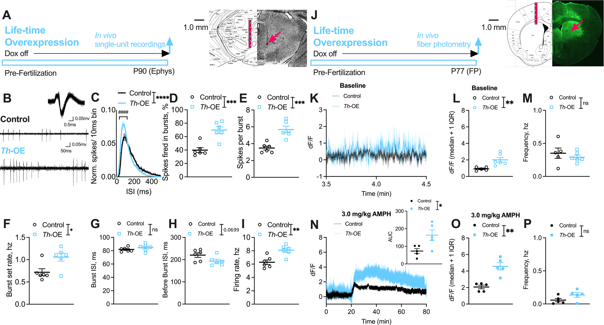
Figure 3. Slc1a1-Th-OE mice show increased dopamine transmission.

(A-I) Single-unit recordings. (A) Representative coronal section showing electrode track (arrow) through the VTA recording region. (B) Representative extracellularly recorded dopaminergic neuron waveform (overlay of ~100 spikes) (Upper) and spiking patterns (Lower) from control and Th-OE mice. For all ephys panels, n = 62 cells/ 6 control mice, 59 cells/ 6 Th-OE mice. (C) Normalized ISI histograms from putative dopaminergic neurons showing increased proportion of short duration ISIs in Th-OE mice. Th-OE mice display increased (D) % spikes fired in bursts, (E) spikes per burst and (F) burst set rate. (G) Average burst ISI is unaltered and (H) average before-burst ISI show trend level decreases in Th-OE mice. (I) Overall firing rate is increased in Th-OE mice. (J-P) dLight1.1 fiber photometry. (J) Representative coronal section showing optic fiber placement and dLight1.1 expression in the dorsal striatum. (K) Dopaminergic transients during baseline evaluation period. For all baseline panels, n = 5 control, 6 Th-OE mice. (L) Th-OE mice show increased fluorescence (median) compared to littermate control mice. (M) No genotype differences were observed in transient frequency. (N) Th-OE mice display increased fluorescence after AMPH challenge in comparison with littermate controls. For all AMPH panels, n = 5 per genotype. (O) Increased dopaminergic transient fluorescence (median), and (P) unaltered transient frequency in Th-OE mice compared to littermate controls. ****P < 0.0001; ***P < 0.001; **P < 0.01; *P < 0.05; ####P < 0.0001; nsP, not significant.
