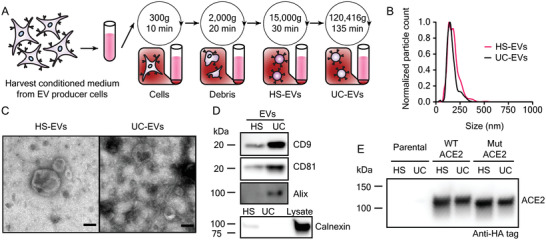
Figure 2.

Extracellular vesicles display classical EV characteristics and EVs from engineered cells contain ACE2. A) Depiction of the process used to isolate extracellular vesicles used in this study. B) Representative histogram of nanoparticle tracking analysis of HEK293FT EV subpopulations normalized to the modal value in each population. C) Transmission electron microscopy images of representative EV subpopulations. Scale bar represents 100 nm. D) Western blots of EVs evaluating standard markers CD9, CD81, and Alix, and a blot of EVs and cell lysate evaluating the potentially contaminating endoplasmic reticulum protein, calnexin (n = 1). E) Western blot against the C‐terminal HA‐tag of transgenic ACE2 in EV populations from parental or engineered cell lines (n = 2). Western blots were normalized by vesicle count (for EVs). The calnexin western blot comparing lysate to EVs used the same number of EVs per well as for CD9, CD81, and Alix and 3 µg cell lysate.
