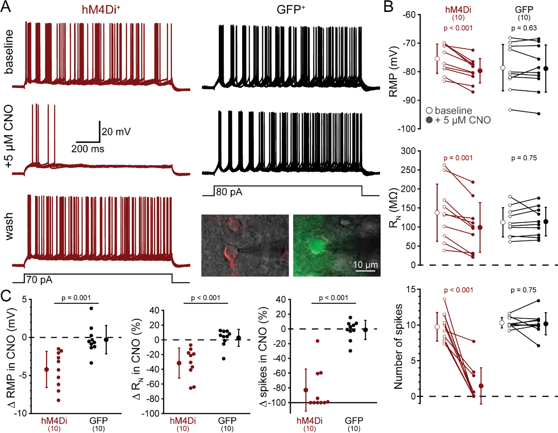
Figure 4.

CNO selectively reduces the excitability of hM4Di-expressing cortical neurons in the postrhinal cortex. A, Ten consecutive voltage traces (superimposed) recorded in response to current injections (black traces at bottom) in a neuron expressing mCherry-tagged hM4Di receptors (red, left) or from a GFP-expressing neuron (black, right) in baseline conditions (top traces) or after 5 minutes exposure to CNO (5 μM). A lower trace for the hM4Di-expressing neuron shows reversal of CNO-induced inhibition following removal of CNO (“wash”). Inset are fluorescence images (artificially colored) of punctate mCherry (left) and diffuse GFP (right) superimposed on oblique images of the two neurons from which traces were taken. B, Measurements of resting membrane potential (RMP, top), input resistance (RN, middle), and number of action potentials (bottom) in baseline conditions (open circles) and after application of 5 μM CNO (filled circles) for ten hM4Di-expressing (red) and ten GFP-expressing (black) neurons. Means (± standard deviations; SD) for each condition indicated by enlarged symbols; p-values from paired Student’s t-tests. C, Comparisons of CNO-induced changes in RMP (left), RN (middle), and action potential number (right) in hM4Di-expressing (red) and GFP-expressing (black) neurons. Means (± SD) for each group indicated by enlarged symbols; p-values from Student’s t-tests comparing results across hM4Di+ and GFP+ neurons.
