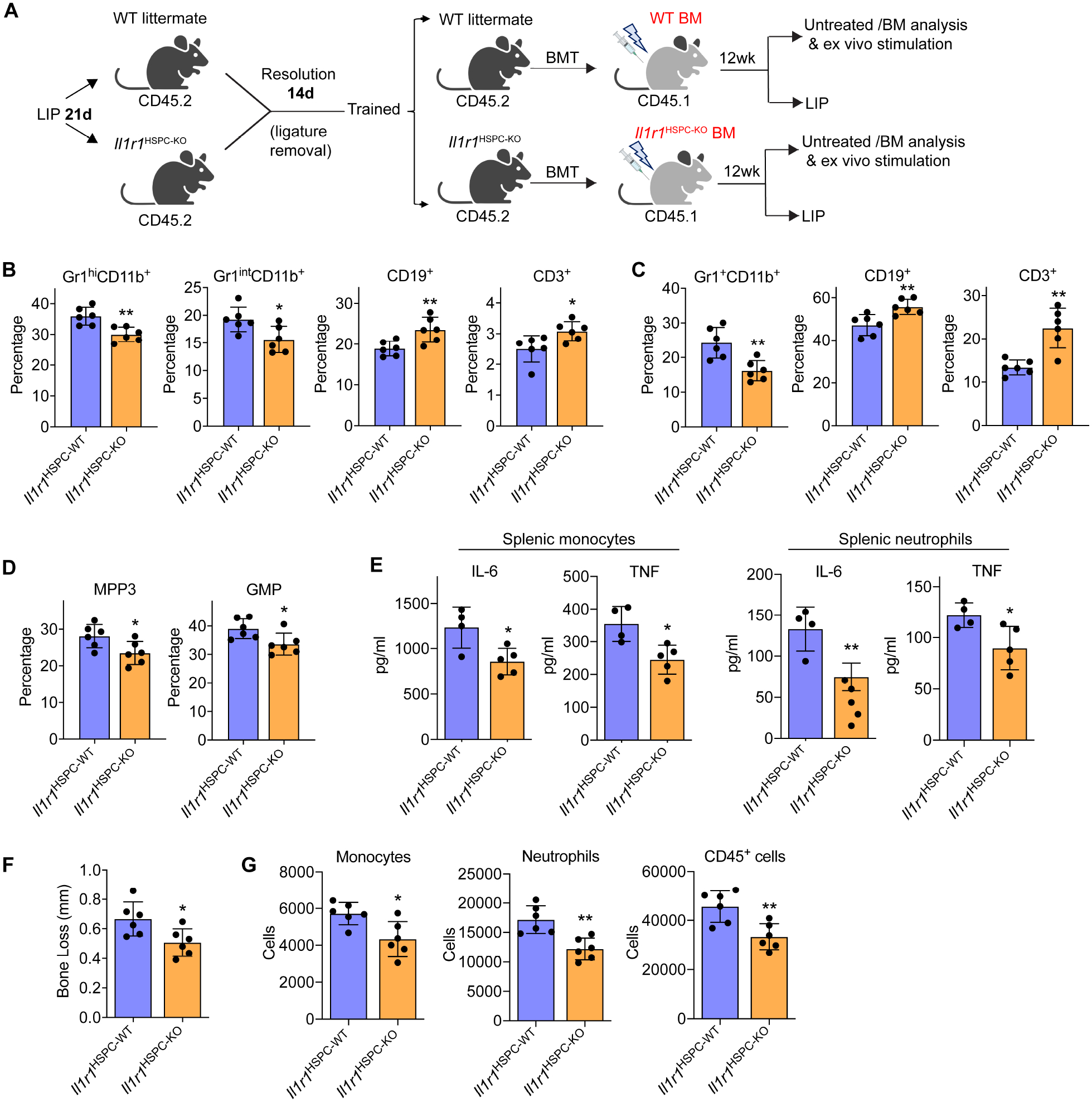
Figure 7. IL-1-signaling in HSPC mediates LIP-induced maladaptive training of myelopoiesis.

(A) CD45.2+ Il1r1HSPC-KO and littermate controls with intact HSPC IL-1R expression were trained (21-day LIP and 14-day resolution) and used as donors for BMT to lethally irradiated congenic B6.SJL (CD45.1+) mice. (B-D) 12 weeks post-BMT, the frequencies of indicated myeloid cells and lymphocytes in CD45+ cells in BM (B) and peripheral blood (C) of recipients, and the frequencies of MPP3 (in LSK) and GMP (in MyP) (D) were determined by FACS. (E) Splenic monocytes (left) and neutrophils (right) isolated from recipient mice were stimulated with LPS (10ng/ml) for 24h and IL-6 and TNF concentrations in the supernatant were measured. (F,G) The recipient mice were subjected to 5-day LIP, as shown in (A) and assayed for bone loss (F) and abundance of gingival monocytes, neutrophils and total CD45+ cells by FACS (G). Data are means±SD (n=4–6 mice/group). *P<0.05, **P<0.01, NS, not significant vs. WT littermate controls; two-tailed Student’s t-test. T, Trained; U, Untrained. See Figure S7.
