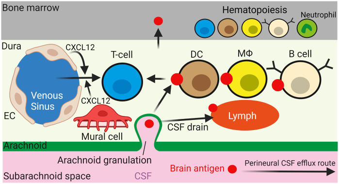
Fig. 3.
Dural immune cells survey brain antigens supplied by the draining CSF. CSF antigens may enter the dura through arachnoid granulations and become captured by APCs including DCs, , and possibly B cells. Endothelial (EC) or mural cell release of CXCL12 recruits T cells across the fenestrated endothelium of dural venous sinuses (on left), allowing T cells to survey antigens presented by APCs. CSF antigens are presented to immune cells in cervical lymph nodes (not shown) by being drained along perineural and dural lymphatic routes. Meningeal immune cells are repopulated by immune cells generated in the skull bone marrow. Figure created using BioRender.54

