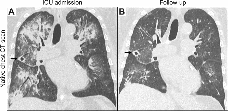
Fig. 1.
Native CT scans of the chest at the time of ICU admission and during follow-up. A At the time of admission, a CT scan confirmed diffuse pulmonary hemorrhage in the upper lobes and right lower lobe with widespread areas of consolidation with surrounding ground glass opacities and crazy-paving as well as a cavitating nodule (arrow) in the middle lobe consistent with GPA. B A follow-up CT scan 2 weeks after ICU admission revealed regression of the diffuse pulmonary hemorrhage with residual linear areas of consolidation and focal ground glass opacities. The cavitating nodule in the middle lobe remained unchanged (arrow). CT computed tomography, GPA granulomatosis with polyangiitis, ICU intensive care unit

