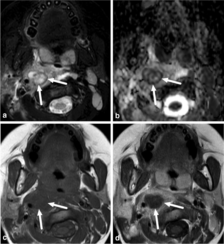
Fig. 4.
Retropharyngeal suppurative lymphadenitis in a 7-year-old girl with a sore throat and streptococcal tonsilitis. a–d Axial images show a focal mass in the right lateral retropharyngeal space (arrows) with an intermediate T2-signal (a), low apparent diffusion coefficient consistent with restricted diffusion (b) and no central contrast enhancement (c T1-weighted, before contrast; d T1-weighted after contrast), consistent with suppurative lymphadenitis. Purulence was found during surgery

