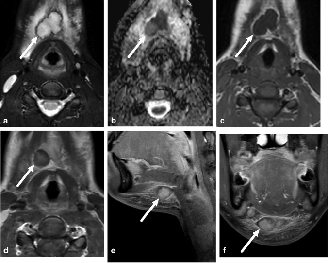
Fig. 6.
False-positive abscess in a 15-year-old girl with neck swelling. a Axial fat-saturated T2-weighted magnetic resonance imaging (MRI) slices (top left) demonstrates lymphadenitis (arrow) and surrounding tissue edema. b Axial apparent diffusion coefficient image shows restricted diffusion in the lesion (arrow). c, d Axial T1-weighted images before (c) and after (d) contrast shows faint partial contrast enhancement compared with pre-contrast image (arrows). e, f Sagittal (e) and coronal (f) fat-saturated post-contrast T1-weighted images confirm faint enhancement (arrows). This finding was interpreted as suppurative lymphadenitis (intranodal abscess). Surgery found necrotic lymphadenitis, but no purulence. This case highlights the difficulties involved in distinguishing necrosis from early abscess

