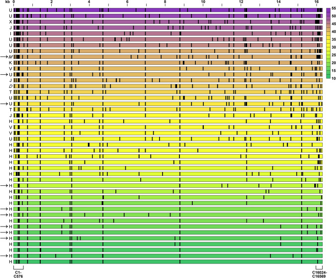
Fig. 1.
Mitochondrial DNA (mtDNA) variant number in whole blood from 44 high-risk rectal cancer patients. Each row represents one individual patient with the haplogroup shown to the left (the non-European M and D haplogroups in italic). The positions across the entire ~ 16.6 kilobase (kb) mitochondrial genome are indicated on the top of the panel. The control (C) region’s base sites 1–576 and 16024–16569 are specified below the panel. Arrows mark patients who had metastatic progression during the follow-up after completion of the curative-intent therapy; one of the metastatic haplogroup U cases was a patient who had declined primary tumour surgery (i.e., refused the full treatment). Each vertical line indicates the position of an mtDNA base with at least one variant other than the reference, as called by the curated reference genome (mitomap.org). The total number of base variants in each case is indicated by colour (scale to the right)

