Fig. 1.
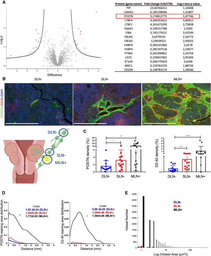
Periostin is upregulated in sentinel LNs from patients with early cervical cancer. a The marginal sinus of LN from patients with a cervical carcinoma has been laser-micro dissected (n ≥ 5). Protein extracts were subjected to proteomic analysis using mass spectrometry. Volcano plot of the proteomic analysis. Volcano plot based on the mean of the protein fold change associated with its P value (− LogP). Each red dot corresponds to a statistically significant protein between the sentinel and the control LN. Proteins modulated between the sentinel and the non-sentinel LN are indicated in the table. b Double immunostaining of POSTN (green), Podoplanin (D2-40 in red) and nuclei (DAPI, blue) on human LNs: distant negative LNs (DLN−, n = 13), negative sentinel LN (SLN−, n = 13) and metastatic LNs (MLN+, n = 12). Scale bars represent 500 µm. c Computer-assisted quantification of POSTN and Podoplanin densities (in percentage) in sentinel and metastatic LNs. Graphs are presented as scatter plots of individual data points. Results are expressed as mean ± SD (Wilcoxon–Mann–Whitney test: p* < 0.05; p** < 0.01; p*** < 0.001; p**** < 0.0001). d Spatial lymphatic vessel and POSTN distribution from tissue edge to tissue center measured on whole tissues of DLN−, SLN− and MLN+ (statistical analyses: Kolmogorov Smirnov test, *p < 0.05). Maximum distance of migration from the tissue border (Lmax) is indicated and expressed as mean ± SD. e Lymphatic vessel size distribution

