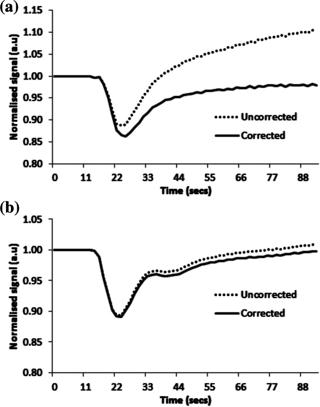
Fig. 4.

Example of signal–time curves from a low-grade tumor and a high-grade tumor, both scanned at center 2 with the same dynamic susceptibility-contrast MRI protocol. No pre-bolus contrast agent was given in either case. The uncorrected and leakage-corrected signal–time curves are shown for both tumors. a The low-grade tumor is a grade I pilocytic astrocytoma in a 2-year-10-month-old boy. Uncorrected and corrected median relative cerebral blood volume (rCBV) for the low-grade tumor are −0.82 and 1.08, respectively. K2 is large and positive at 0.022. b The high-grade tumor is a grade IV medulloblastoma in a 5-year-5-month-old boy. Uncorrected and corrected median rCBV for the high-grade tumor are 1.30 and 1.23, respectively. K2 is low at 0.001
