
Figure 3.
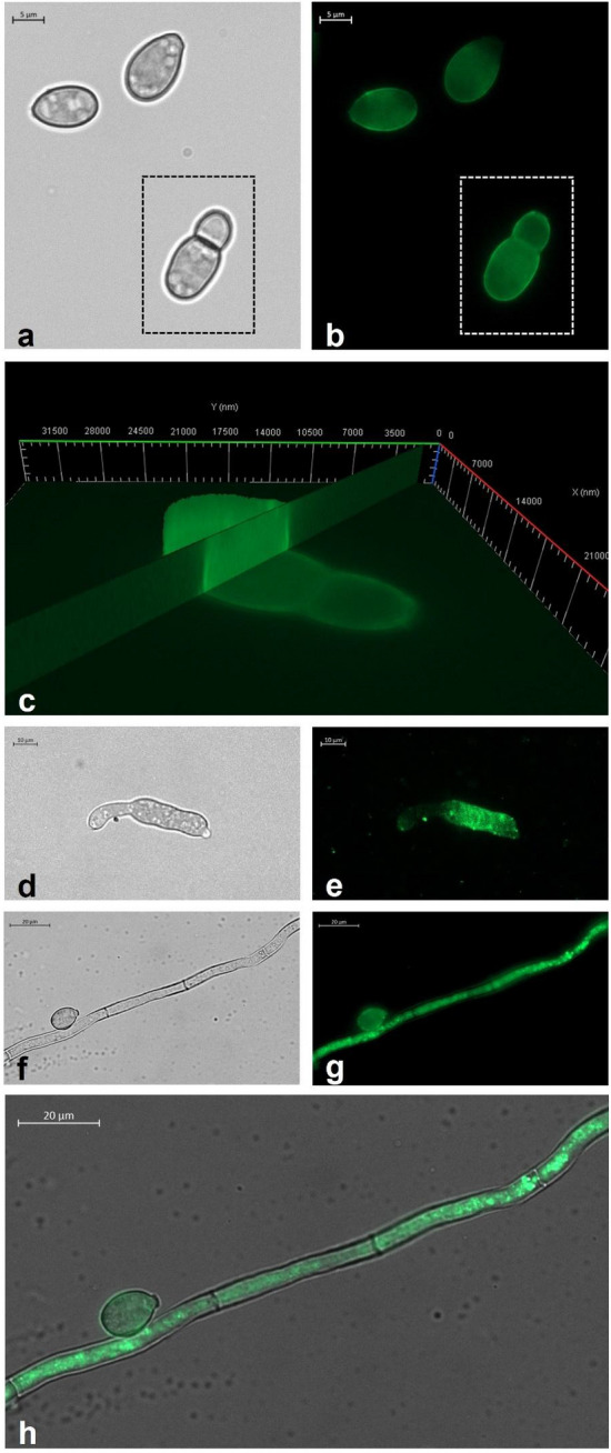
(a) Bright field and (b) fluorescence image of B. cinerea conidia treated for 10 min with Cu6-PLGA NPs. NPs fluorescence signal was evident in germinated B. cinerea conidia (white square) and non-germinated ones. (c) 3D reconstruction of B. cinerea conidia treated with NPs for 10 min. The fluorescence signal was detected inside conidia, along the whole thickness of the spore. (d) Bright field and (e) fluorescence image of B. cinerea germ tube. The NPs fluorescence signal was well visible in the expanding germ tube after 10 min of administration. (f) Bright field and (g) fluorescence image of B. cinerea conidium and hypha. The fluorescence signal was well visible both in the conidium and the hypha after 1 h of administration. (h) Overlap of the bright field image and the fluorescence image which shows the localization of Cu6-PLGA NPs inside the fungal conidium and hypha.