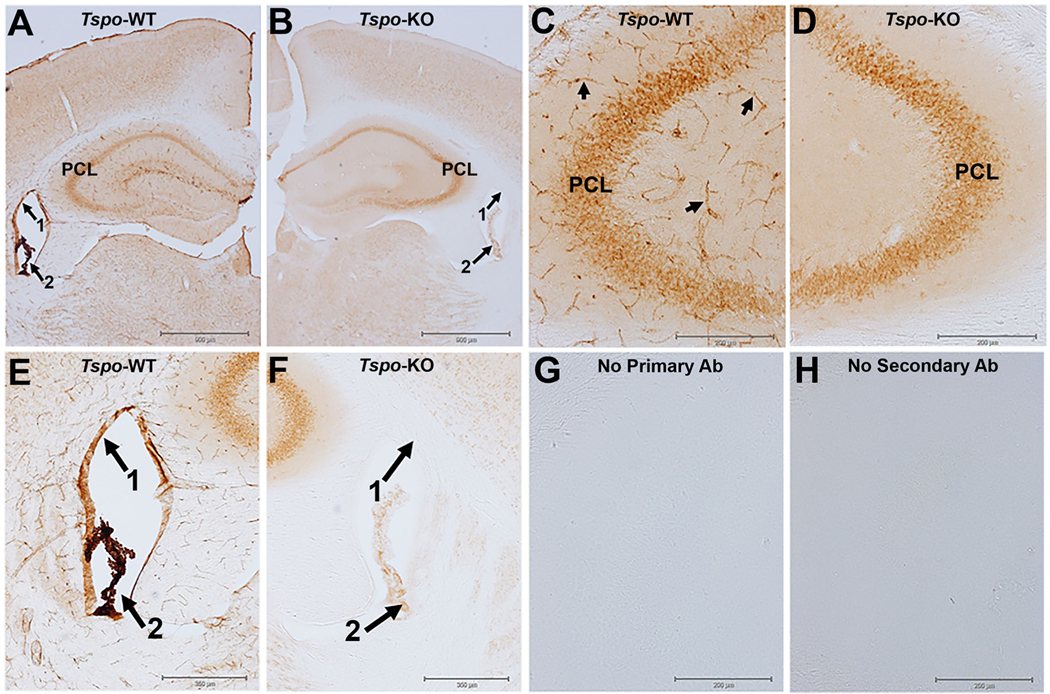
Fig. 1.

TSPO immunohistochemistry with diaminobenzidine (DAB) visualization in the hippocampus of wildtype and TSPO global knockout mice.
A) TSPO immunostaining in a normal wildtype (WT) animal at the level of the hippocampus with high expression of TSPO in the ependymal cells of the lining of the ventricles (arrow 1), in the choroid plexus (arrow 2), in blood vessels throughout the brain neuropil, and in pyramidal neurons (PCL) of the hippocampus. B) Image of TSPO immunostaining at the same level of the hippocampus as in panel A but the brain tissue is from a global TSPO-KO mouse that was processed at the same time and under the same experimental conditions as in panel A. The image in the TSPO-KO tissue shows the complete loss of TSPO staining in the ependymal cells of the ventricles, blood vessels, and a nearly complete loss in the choroid plexus. However, there was essentially no loss in pyramidal neurons (PCL) of the hippocampus indicative of non-specific TSPO staining since the tissue is from a TSPO-KO mouse. C and D) are higher magnification images at the level of the CA3 region of the hippocampus. Arrows in Panel C point to TSPO staining of blood vessels in the WT tissue with the TSPO staining disappearing in blood vessels of the TSPO-KO mouse. E and F) are higher magnification images of the lateral ventricles. G and H) are images from the no primary or no secondary antibody controls. Brain slices from wildtype (WT) and TSPO knockout (KO) mice were processed using standard methods with the TSPO primary antibody (ab109497 Abcam) which has been extensively validated using multiple approaches including TSPO-KO cells and tissue (Loth et al., 2020 and supplementary materials within). Experimental methods are described in supplementary materials section.
