
Fig. 2.
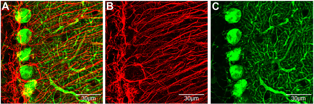
Immunofluorescent confocal imaging of Purkinje cells and Bergmann glia in the cerebellar cortex.
A) Top-view of a 3D condensed z-stack of double label immunofluorescence confocal imaging of Purkinje cells (calbindin-green) and Bergmann glia (GFAP-red). B and C) are the presentation of the individual channels. Bergmann glia cell bodies closely contact and wrap Purkinje cell soma and processes. One distinguishing anatomical feature of Bergmann glia is that they have processes that originate at their soma in the Purkinje cell layer and have a relatively straight and lengthy trajectory to the pial surface of the molecular layer (Reeber et al., 2018). On the other hand, Purkinje cell processes are shorter and radiate at all angles throughout the molecular layer. Green = Purkinje cell; Red = Bergmann glia. (For interpretation of the references to colour in this figure legend, the reader is referred to the web version of this article.)