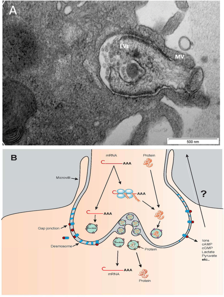
Fig. 1.
A Electron microscopy image of a bovine TZP terminus, the bulged portion held in place by microvilli (MV). Numerous extracellular vesicles (EVs, spheres about 50 nm in diameter) are visible. The projection terminus contains electron-dense structures. B Schematic representation of material movement through a TZP terminus. Transcripts (mRNA) could be transferred to the oocyte or translated polyribosomes, and proteins could be transferred. Small molecules could be transferred through gap junctions

