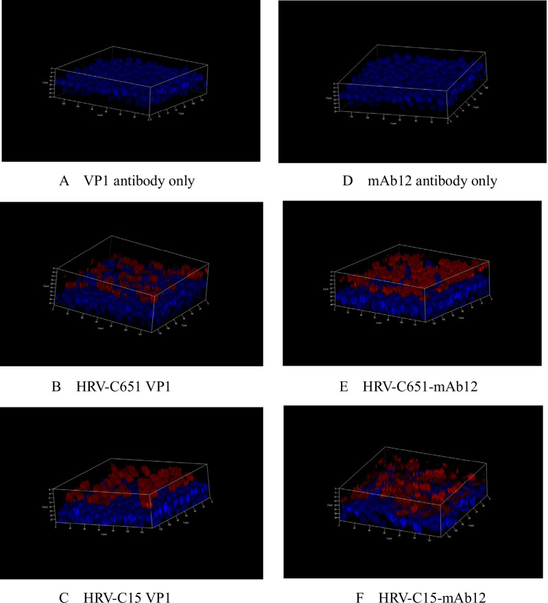
Fig. 2.
Immunofluorescence of differentiated HBE cells infected with HRV-C651 or HRV-C15. Immunofluorescence with the rabbit polyclonal anti-HRV-C VP1 protein-antibody only (A) and mAbJ2 antibody (D). At 24 h post-inoculation with HRV-C651 or HRV-C15, the HBE cells were fixed, followed by incubation with VP1 protein-antibody (B and C) and mAb12 antibody detecting double-strand RNA (E and F). The red region indicates VP1 protein (B and C) or the double-stranded RNA of HRV-C (E and F). Confocal images were taken with a magnification of 200×. Nuclei were stained with DAPI

