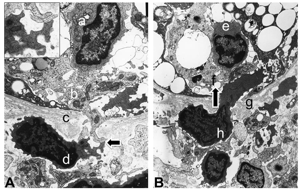
Figure 2. Interaction of acute cellular rejection captured by electron microscopy.

Panel A (left). Leaked proximal tubular cytoplasm, through tubular basement membranes interacts with extracellular materials of lymphocytes as an induction of lymphocytic infiltration. Letters represent following structures: a. an infiltrated lymphocyte in the proximal tubule (tubulitis); b. cytoplasm of proximal tubules with leaking component through tubular basement membrane (c); d. lymphocyte releasing extracellular particles to interact with proximal tubular cytoplasm (indicated by an arrow; details presented in the left upper corner insert). Panel B. Activated lymphocyte physically penetrates through tubular basement membrane into the proximal tubule (infiltration direction is indiacted by an arrow). Letters represent following structures: e. infiltrated lymphocyte (tubulitis); f. proximal tubular epithelium; g. tubular basement membrane; h. lymphocyte extending beyond tubular basement membrane as an action of infiltration.
