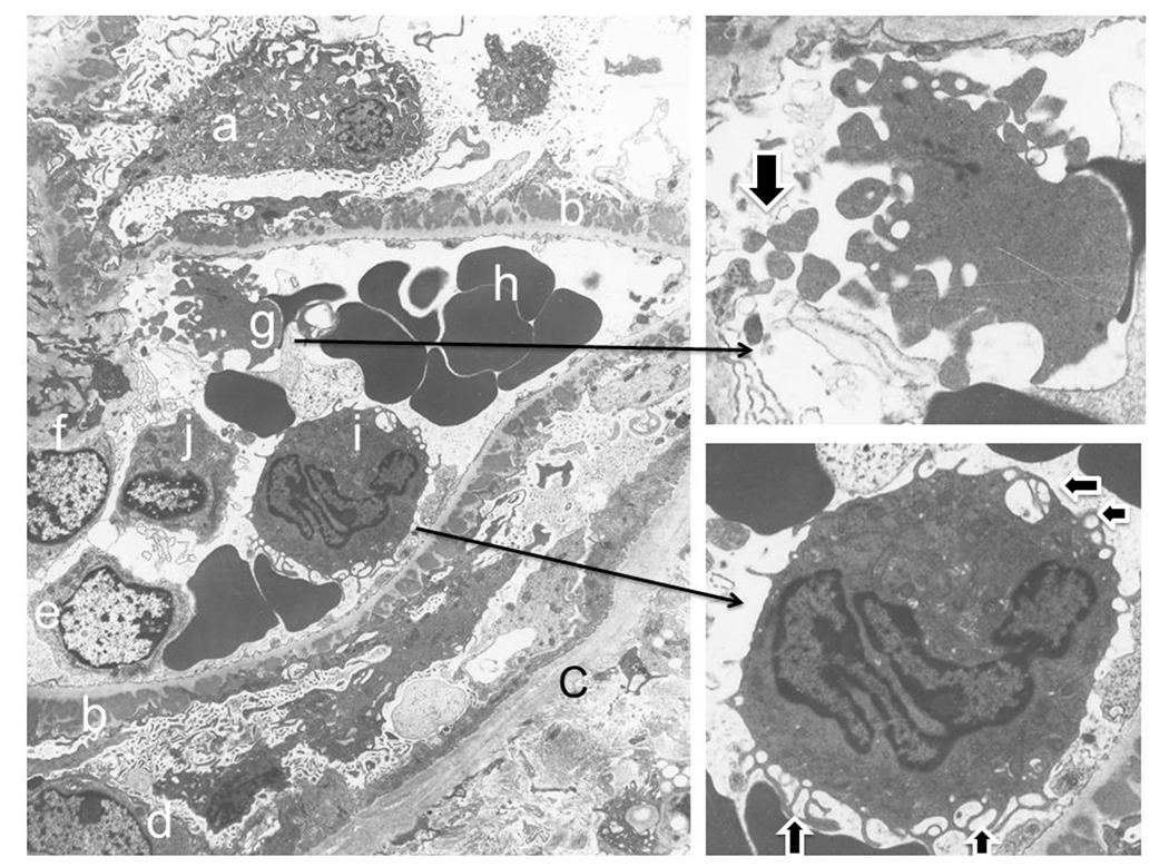
Figure 3. Shedding platelet fragments and releasing extracellular vesicles of neutrophil as early sign of thrombosis in membranous glomerulopathy.

The left large panel of image captures shedding platelet fragments with a glomerular capillary loop, which details can be further seen in the right upper insert (fragments indicated by an arrow). Meanwhile, an activated neutrophil within the same loop demonstrates its exosome vesicles in the cytoplasm surface (indicated by two horizontal arrows in right lower panel insert) and releasing exosomes and/or microparticles from its cell surface membranes (two vertical arrows in the right lower panel inset). Letters represent following structures: a. podocyte; b. subpeithelial immune complex deposits above the glomerular basement membrane; c. Bowman’s capsule, d, parietal epithelial cell; e. glomerular endothelial cell; f. mesangial cell; g. platelet with fragmentation (also see in the insert in right upper panel); h. red blood cells; i. activated neutrophil (also see in the insert in right lower panel); j. lymphocyte.
