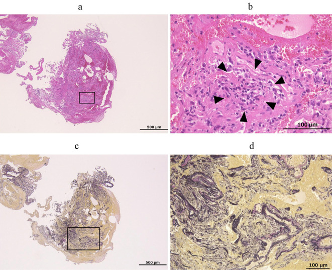
Figure 2.
Histological findings of the transbronchial lung biopsy specimen. A histological examination was performed with Hematoxylin and Eosin staining (a, b) and the Elastica van Gieson staining (c, d). The square area of the low-magnification photomicrograph (a, c) is shown as a high-magnification photomicrograph (b, d) with each stain. The arrowheads indicate the infiltration of lymphocyte into the alveolar interstitium. A scale bar is shown in each figure.

