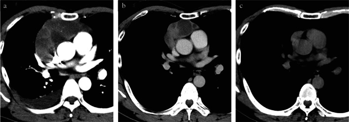
FIGURE 1.

Computed tomography findings. (a) At initial visit: an anterior 11.0 cm mediastinal mass with right pleural effusion. (b) After 1 year of follow‐up: the anterior mediastinal mass has spontaneously regressed to 5.5 cm. The right pleural effusion has disappeared. (c) Four years after the initial visit: the mass has regressed to 3.0 cm. The cystic component is obscure
