
FIGURE 3.
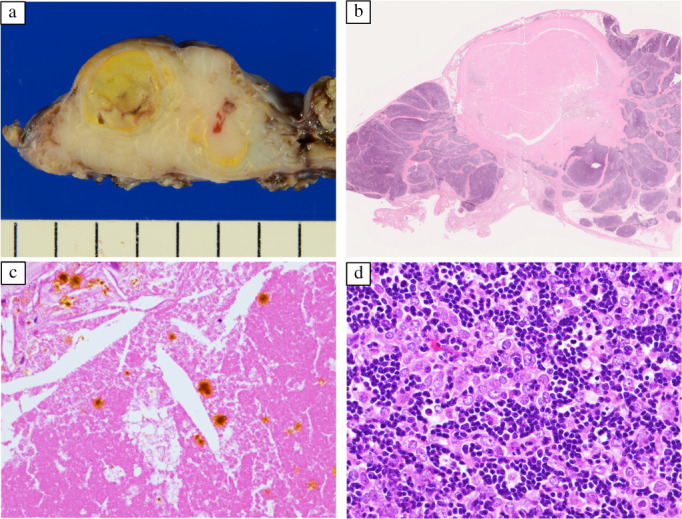
Pathology findings. (a) The tumor is encapsulated, with a white cut surface and a well‐circumscribed yellowish‐white internal area. (b) A low‐power view: the tumor contains a broad necrotic area in the center (hematoxylin–eosin staining). (c) A high‐power view: cholesterol clefts and hematoidin deposits are present in necrotic areas (hematoxylin–eosin staining). (d) A high‐power view: polygonal epithelial cells and abundant lymphocytes suggest thymoma type B2 (hematoxylin–eosin staining)