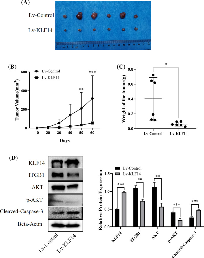
Fig. 3.
KLF14 inhibited the progression of cervical cancer in vivo. A First, six groups were set up. The injection scheme was as follows: six nude mice were used, and the left and right sides of each nude mouse were compared. SiHa cells (5 × 10⁶ cells/group) of the Lv-KLF14 group were injected subcutaneously into the axilla of the right upper limb of nude mice with 200 μl PBS suspension as the positive control. The Lv-control group was injected subcutaneously into the left upper arm axilla of nude mice with 200 μl PBS suspension as the negative control. Next, DOX, as an inducer, induced KLF14 expression in the Lv-KLF14 group, while KLF14 was not expressed in the Lv-control group, allowing for comparison. The induction scheme was as follows: before the injection, SiHa cells of the Lv-KLF14 group and Lv-control group were cultured in medium containing DOX (5 µg/ml) for 48 h. After the injection, the nude mice were fed water with DOX (1 mg/ml) for 1 week. After that, DOX-containing water was provided intermittently. Two months later, the nude mice were sacrificed, and the size of subcutaneous tumours in the Lv-KLF14 group was smaller than that in the Lv-control group. B Tumour growth was recorded every 10 days, and tumour volume was calculated using the formula V = (larger diameter) × (smaller diameter)2/2. The results showed that tumour growth was faster in the Lv-control group than in the Lv-KLF14 group. C The weight of subcutaneous tumours in the Lv-KLF14 group was lower than that in the Lv-control group. D According to the Western blot results, the expression of KLF14 and cleaved caspase-3 was upregulated and the expression of ITGB1 and p-AKT was downregulated in the Lv-KLF14 group compared with the Lv-control group in tumour tissues. (*P < 0.05, **P < 0.01, ***P < 0.001)

