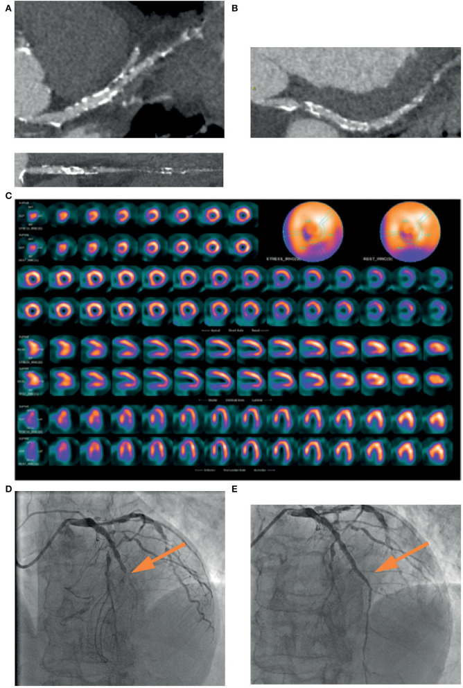
Figure 2.
A 74-years-old patient, symptomatic for chest pain, performed a coronary CT, which showed a mild stenosis of left main and a diffuse severe stenosis of mid-distal left anterior descending (LAD) coronary artery (A). Circumflex artery had a significant ostial stenosis, with sub-occlusion of mid-distal portion (B) and right artery was occluded at the mid portion (C). SPECT showed perfusion defect at the apical septum and at infero-lateral mid-basal wall (D). Coronary angiography confirmed the presence of sub-occlusion (yellow arrow) of the middle section of the LAD coronary artery (E), treated with coronary angioplasty and placement of a medicated stent (yellow arrow), with a good final angiographic result (F).

