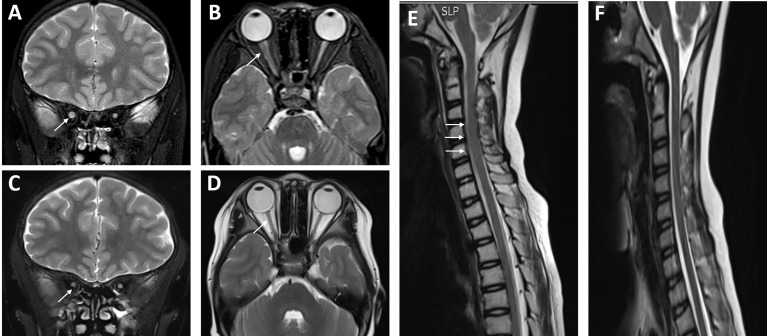
Figure 3.
Neuroimaging changes of the patient with systemic lupus erythematosus (SLE)/Sjogren’s syndrome (SS) and NMOSD. (A, B) The left optic nerve was obviously smaller than the right through MRI, and there was slight swelling of the spinal cord at the levels of c2-6 (E) in December 2018 (shown by arrows). After a powerful immunosuppressive therapy, the left optic nerve was roughly the same size as the right optic nerve in October 2019 (C, D), and the swelling of c2-6 was much better than before (F).

