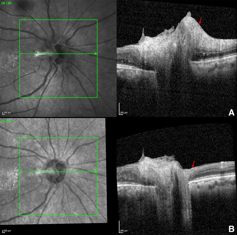
Figure 6.
Spectral domain optical coherence tomography image of retinal nerve fibre layer of right eye showing (A) peripapillary swelling (red arrow) with hyperreflective band into vitreous cavity correlating with the fibrous remnant at 8 months earlier, (B) significant reduction in peripapillary swelling (red arrow) at presentation now.

