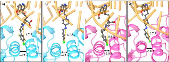
Figure 6.
Comparisons of the binding pocket sizes of S. aureus DNA gyrase and topoIV as well as docking-derived poses of compounds 45 and 46, with different RHS fragment lengths. (a) S. aureus DNA gyrase crystal structure (cyan, PDB ID: 6Z1A)22 with docked pose of 45; (b) S. aureus DNA gyrase crystal structure with docked pose of 46; (c) S. aureus topoIV homology model (pink) with docked pose of 45; and (d) S. aureus topoIV homology model with docked pose of 46. The enzymes are represented as cartoons, and compounds as balls and sticks, while DNA is in orange.

