Figure 2.
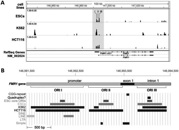
Sites of replication initiation in the vicinity of the FMR1 gene in a panel of human cell lines. Panel (A) shows the nascent strand abundance in the 100 kb region containing FMR1 in ESCs, K562 and HCT116 cells as determined by deep sequencing of nascent strands. The nascent strand peaks at the 5′ end of the FMR1 gene are shown in the shaded block. This block, which spans ∼3000 bp, contains three regions that show evidence of ORI activity in some or all of the cell lines tested. These regions are designated as ORI I, II and III. Panel (B) shows an expanded view of the shaded region shown in (A) illustrating the position of the three ORIs along with various sequence elements present in this region including the CGG repeat, a potential quadruplex and the remnants of two long interspersed repeated DNA (LINE) elements. The ORI cores, defined as those regions showing the highest number of nascent DNA strands in deep sequencing, are also shown. In both (A and B), the numbering refers to the nucleotide numbering on the X chromosome from the GRCh37/hg19 assembly of the human genome.

