Figure 4.

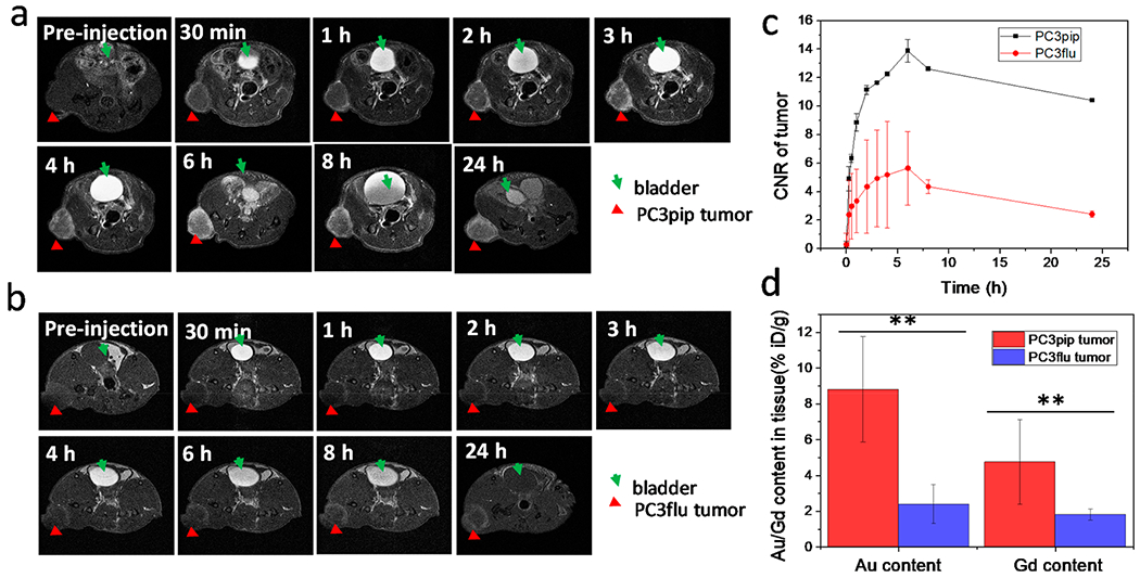
In vivo tumor targeting of Au–Gd(III)-PSMA NPs and MR imaging. T1-weighted spin echo images of mice with PC3pip tumor (a) and PC3flu tumor (b) obtained at 7 T. Tumors are indicated by red triangles and bladders are indicated by green arrows. Representative images are shown of n = 3. (c) Contrast-to-noise ratio (CNR) of PC3pip and PC3flu tumors relative to muscle, computed from T1-weighted images (n = 3). (d) Au and Gd(III) content in PC3pip and PC3flu tumors 24 h after Au–Gd(III)-PSMA NPs injection. Data are presented as the mean ± SD (n = 3), and differences between groups are compared with two-tailed t tests, **p ≤ 0.01.
