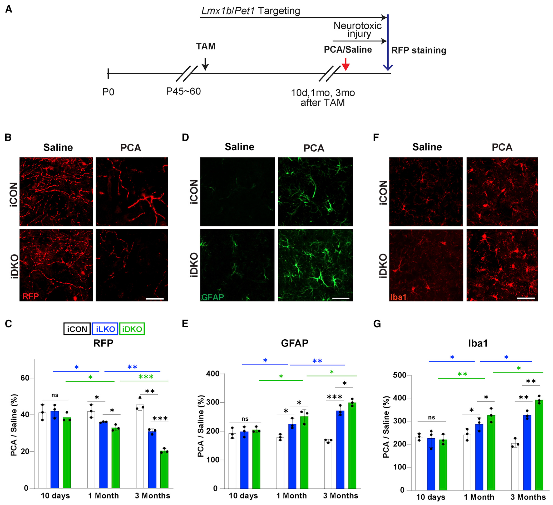
Figure 5. Adult-stage targeting of Lmx1b and Pet1 causes increased susceptibility of 5-HT axons to neurotoxic injury.

(A) Experimental design.
(B and C) Representative images at 1 month post-TAM (B) and quantification (C) of anti-RFP fluorescence intensities at indicated times post-TAM shows greater loss of TdTomato+ axons in somatosensory cortex of iLKO and iDKO versus iCON mice treated with PCA. Presented as ratio of TdTomato+ fiber intensities in PCA versus saline treated mice.
(D and E) Representative images of GFAP immunopositive cells (D) and quantification of immunofluorescence intensities (E) in somatosensory cortex at 1 month post-TAM.
(F and G) Representative images of Iba1 immunopositive cells (F) and quantification of immunofluorescence intensities (G) in somatosensory cortex at 1 month post-TAM. two-way ANOVA; ±SEM; n = 3 mice per genotype. *p < 0.05, **p < 0.01, ***p < 0.001. ns, not significant. Scale bars, 5μm.
